|
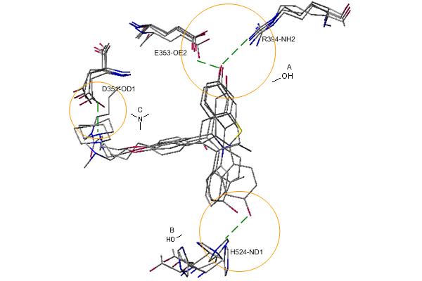
Figure 2.
Binding-site pharmacophores identified by superimposing four crystal structures of ERα shown in Figure 1. Three pharmacological preferences and interactions are identified and circled as A ( a hydroxyl group ), B ( a basic group ), and C ( a hydroxyl group ). The dash lines indicate the hydrogen bonds formed between protein and ligand.
|