|
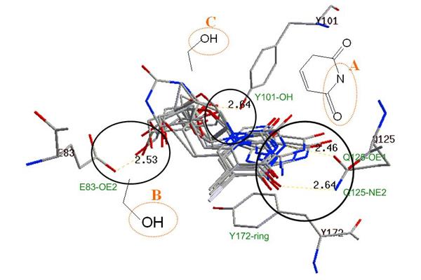
Figure 2. Binding-site phamacophores identified by superimposing ten crystal structures of HSV-1 thymidine kinase shown in Figure 1. Three pharmacological references and interactions are identified and circled as A (an amide binding site), B (a hydroxyl binding site), and C (a hydroxyl binding site). A stack force binding area (Y172-ring) is also indicated. The dash lines indicate the hydrogen binding.
|